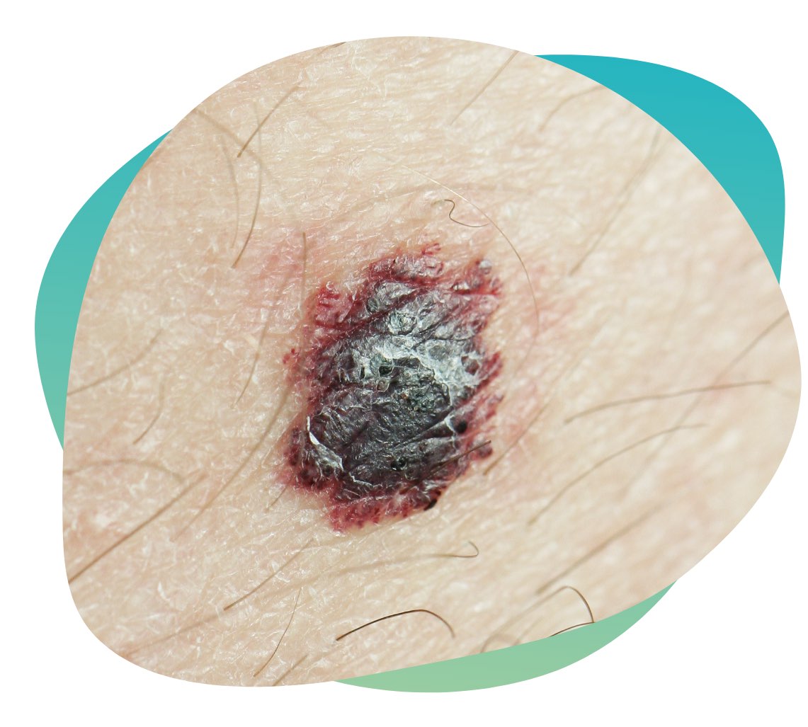
What is Skin Cancer?
Skin cancers are more common than all other cancers combined, and, collectively, their incidence is rising faster than that of any other cancer.

Commonest Cancer Worldwide
1 in 5 people in the US will develop skin cancer by age 70
Melanoma is Deadly Serious
5+ sunburns doubles your risk of getting melanoma
Early Identification is Critical
98+% survival at 5 years if detected whilst localised
Basic Types
Skin cancer may form in basal cells or squamous cells. Basal cell carcinoma and squamous cell carcinoma are the most common types of skin cancer. They are also called nonmelanoma skin cancer. Actinic keratosis is a skin condition that sometimes becomes squamous cell carcinoma.
Melanoma is less common than basal cell carcinoma or squamous cell carcinoma. It is more likely to invade nearby tissues and spread to other parts of the body.

Skin Cancer in the Cayman Islands
In the Cayman Islands, we are blessed with an amazingly sunny climate and a healthy love of the outdoors. Unfortunately, that also comes with an elevated risk of skin cancer. It’s vital to protect from the sun’s harmful UV rays using appropriate clothing and high factor sun creams.
It is also extremely important to remain vigilant for the signs of skin cancer and consider being screened e.g. an annual body map.
As a small island with limited facilities for advanced cancer treatments, the difference between early identification and late is having a local, simple excision of a lesion versus an extended stay at a cancer centre in say Florida. It pays in every way to identify skin cancer early.
Risk Factors
The following is a non-exhaustive list of factors that may increase someone’s likelihood of developing skin cancer (Ref. Mayo Clinic)
- Fair skin
- A history of sunburns
- Excessive sun exposure
- Sunny or high-altitude climates
- Moles
- Precancerous skin lesions
- Family history of skin cancer
- Personal history of skin cancer
- Weakened immune system
- Exposure to radiation
- Exposure to certain substances
The presence of these risk factors heightens the need for vigilance and may warrant a more frequent schedule of screening or follow up.
Symptoms
Melanoma can develop anywhere on your body, in otherwise normal skin or in an existing mole that becomes cancerous. 70+% of melanoma is ‘de novo’ not associated with a nevus (mole). It most often appears on the face or the trunk of affected men. In women, this type of cancer most often develops on the lower legs. In both men and women, melanoma can occur on skin that hasn’t been exposed to the sun.
Melanoma can affect people of any skin tone. In people with darker skin tones, melanoma tends to occur on the palms or soles, or under the fingernails or toenails.
Melanoma signs include:
- A large brownish spot with darker speckles
- A mole that changes in color, size or feel or that bleeds
- A small lesion with an irregular border and portions that appear red, pink, white, blue or blue-black
- A painful lesion that itches or burns
- Dark lesions on your palms, soles, fingertips or toes, or on mucous membranes lining your mouth, nose, vagina or anus
If you have these risk factors, it may be a good idea to book a screening.
If you have active symptoms described above, it is important to consult your dermatologist as early as possible.
Screening
Historically, this involved a dermatologist painstakingly examining and photographing each mole and assigning it to part of the body on a computer. However, with advances in imaging technology and the use of artificial intelligence (AI), this can now be done in just a few minutes. Our state of the art Body Mapping System then analyses the images for suspect areas and stores the images for future reference and change-analysis when you return for the next scan.
Methods of Detection
Once our system has identified lesions or areas of skin that warrant further investigation, these are then examined by our experienced dermatologist, Dr Alison Duncan. She will use high magnification dermoscopy to look at the skin and structures of moles. You may then be scheduled for a follow up examination, have a biopsy taken for analysis, or booked to have the mole removed.